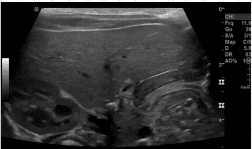
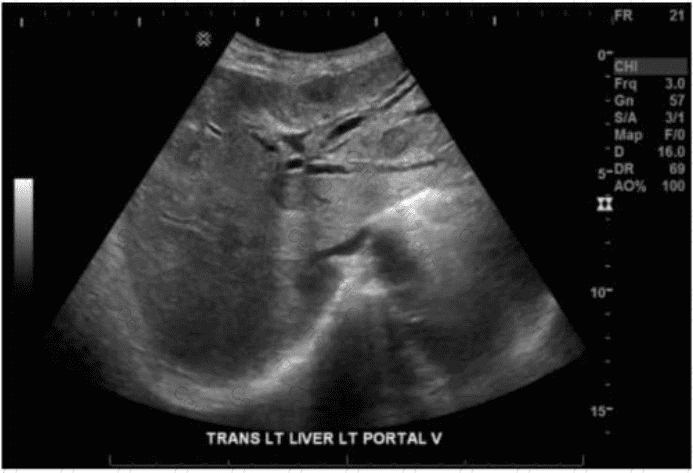
The CT and ultrasound images show a lenticular-shaped, hypoechoic fluid collection compressing the renal parenchyma from the periphery. The location—between the renal capsule and the renal parenchyma—strongly suggests a subcapsular hematoma.
Subcapsular hematomas in transplant kidneys are uncommon but important postoperative complications, often resulting from trauma, biopsy, or surgical manipulation. They typically present as a crescent-shaped, low-attenuation collection beneath the renal capsule and may cause mass effect on the underlying renal parenchyma. This can impair renal perfusion and function—a phenomenon known as “Page kidney.”
Ultrasound findings:
Anechoic to hypoechoic, crescent-shaped collection surrounding or compressing the transplant kidney.
Deformation or flattening of the renal contour due to the compressive effect of the hematoma.
CT findings:
Hyperdense collection (if acute) or hypodense (if subacute/chronic) located subcapsularly.
May be seen distorting the renal capsule and compressing renal tissue.
Differentiation from other options:
A. Urinoma: Typically appears as a perinephric fluid collection and may result from urine leak. Not usually subcapsular.
B. Lymphocele: Develops in the post-transplant period due to lymphatic disruption. Appears as a well-defined perinephric fluid collection and tends to be medial to the kidney or between the bladder and the kidney.
C. Renal cell carcinoma: Would appear as a solid mass with variable enhancement and not a simple fluid collection. RCC is rare in transplant kidneys and usually detected much later post-transplant.
[References:, Radiopaedia.org. Subcapsular renal hematoma. https://radiopaedia.org/articles/subcapsular-renal-haematoma, Rumack CM, Wilson SR, Charboneau JW, Levine D. Diagnostic Ultrasound. 5th Edition. Elsevier, 2018. Chapter: Kidney Transplant Imaging, pp. 524–530., American Institute of Ultrasound in Medicine (AIUM) Practice Parameter for the Performance of an Ultrasound Examination of the Abdomen and/or Retroperitoneum. 2020., , ]