Weekend Sale 70% Discount Offer - Ends in 0d 00h 00m 00s - Coupon code: save70

Free and Premium ARDMS AB-Abdomen Dumps Questions Answers

Page: 1 / 12
Total 165 questions

Abdomen Sonography Examination Questions and Answers

Question 1

Which syndrome is characterized by right upper quadrant pain, ascites, and hepatocellular dysfunction?

Options:

A.

Budd-Chiari

B.

Calciphylaxis

C.

Ehlers-Danlos

D.

Klippel-Trenaunay

Buy Now
Question 2

Which technique best differentiates a bladder mass from a hematoma?

Options:

A.

Use harmonic imaging

B.

Obtain post-void image

C.

Fill the bladder

D.

Change patient position

Question 3

Which portion of the biliary system is last to become dilated with biliary obstruction at the ampulla of Vater?

Options:

A.

Cystic duct

B.

Common bile duct

C.

Common hepatic duct

D.

Peripheral bile ducts

Question 4

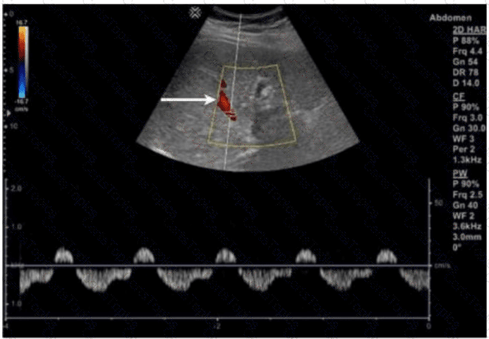
Which portion of the renal arterial vasculature is indicated by the arrow in this image?

Options:

A.

Interlobular

B.

Arcuate

C.

Segmental

D.

Interlobar

Question 5

A patient with hepatocellular carcinoma presents for a paracentesis. Which lab value is the most pertinent to the procedure?

Options:

A.

International normalized ratio

B.

Alanine aminotransferase

C.

Alpha fetoprotein

D.

Total bilirubin

Question 6

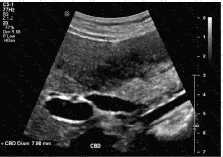
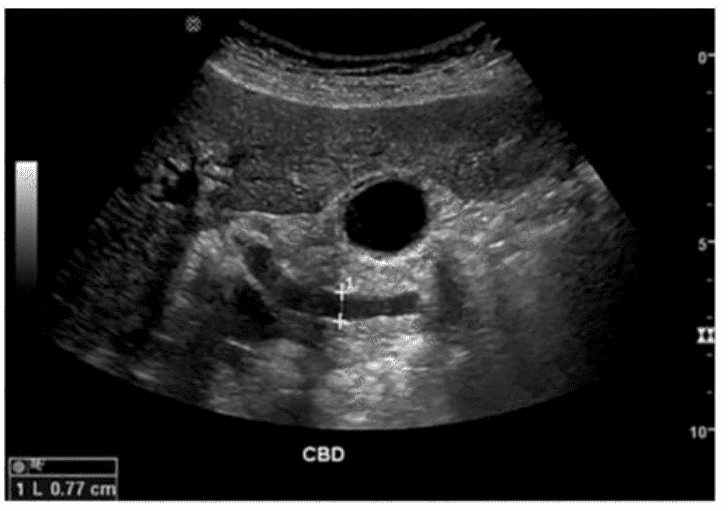
Which term best describes the common bile duct measured in this image of a postcholecystectomy patient?

Options:

A.

Normal

B.

Dilated

C.

Inflamed

D.

Atretic

Question 7

Which condition is demonstrated in this image?

Options:

A.

Cavernous transformation

B.

Portal vein thrombosis

C.

Portal hypertension

D.

Tumor extension

Question 8

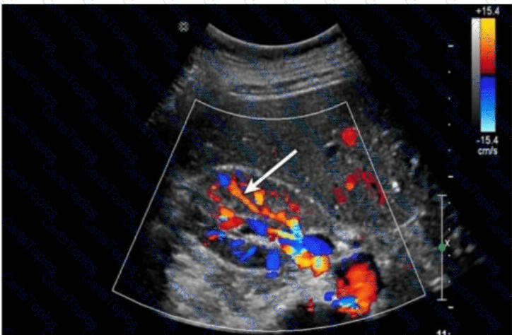
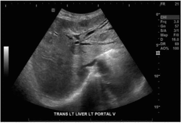
Which sections of the liver are divided by the structure indicated by the arrow on this image?

Options:

A.

Right anterior and posterior

B.

Right and left lobes

C.

Left medial and lateral

D.

Left superior and inferior

Question 9

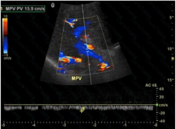
What is the normal Doppler waveform signature of the hepatic veins?

Options:

A.

Low resistant

B.

Monophasic

C.

Triphasic

D.

Turbulent

Question 10

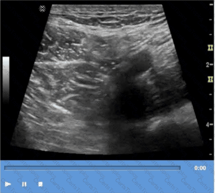
Which technique is used to demonstrate the finding in this video?

Options:

A.

Compression

B.

Valsalva

C.

Exhalation

D.

Deep inspiration

Question 11

Which condition is most likely in a patient presenting with weight loss and fatigue along with elevated liver enzymes, elevated potassium, and decreased sodium?

Options:

A.

Addison disease

B.

Conn syndrome

C.

Acute pancreatitis

D.

Hepatocellular carcinoma

Question 12

Which probe frequency is most appropriate for imaging of the salivary glands?

Options:

A.

2 MHz

B.

4 MHz

C.

8 MHz

D.

12 MHz

Question 13

Which condition is a cause of intrahepatic dilatation with a normal common bile duct?

Options:

A.

Portal vein thrombus

B.

Tumor at the porta hepatis

C.

Choledocholithiasis

D.

Acute pancreatitis

Question 14

Which condition is associated with multiple pancreatic cysts?

Options:

A.

Cystic fibrosis

B.

Von Hippel Lindau syndrome

C.

Beckwith Wiedemann syndrome

D.

Autosomal recessive polycystic kidney disease

Question 15

Which technique is best for demonstrating the characteristic of the small hepatic lesion identified by the arrow on this image?

Options:

A.

Decrease depth

B.

Scan in upright position

C.

Move the transducer focus

D.

Use a standoff pad

Question 16

Which common congenital anomaly is typically seen as a cystic midline anterior neck structure?

Options:

A.

Branchial cleft cyst

B.

Cystic hygroma

C.

Thyroglossal duct cyst

D.

Cystic follicular adenoma

Question 17

Which sonographic finding distinguishes focal nodular hyperplasia from hepatic adenoma?

Options:

A.

Stellate area within the central portion of the mass

B.

Thin peripheral hypoechoic halo

C.

Central calcifications

D.

Target pattern

Question 18

Which sonographic finding is most consistent with scrotal inflammation?

Options:

A.

Abscess

B.

Hydrocele

C.

Granuloma

D.

Hyperemia

Question 19

Which scanning technique is most beneficial when imaging the appendix?

Options:

A.

Apply graded compression around the patient's area of pain

B.

Apply light pressure around the patient's area of pain

C.

Image in the longitudinal plane around the iliac vessels

D.

Image small bowel transversely to evaluate for peristalsis

Question 20

Which of the following would optimize visualization of a bladder mass?

Options:

A.

Compression technique to move bowel gas

B.

Utilize a high-frequency linear transducer

C.

Have patient empty bladder

D.

Have patient fill bladder

Question 21

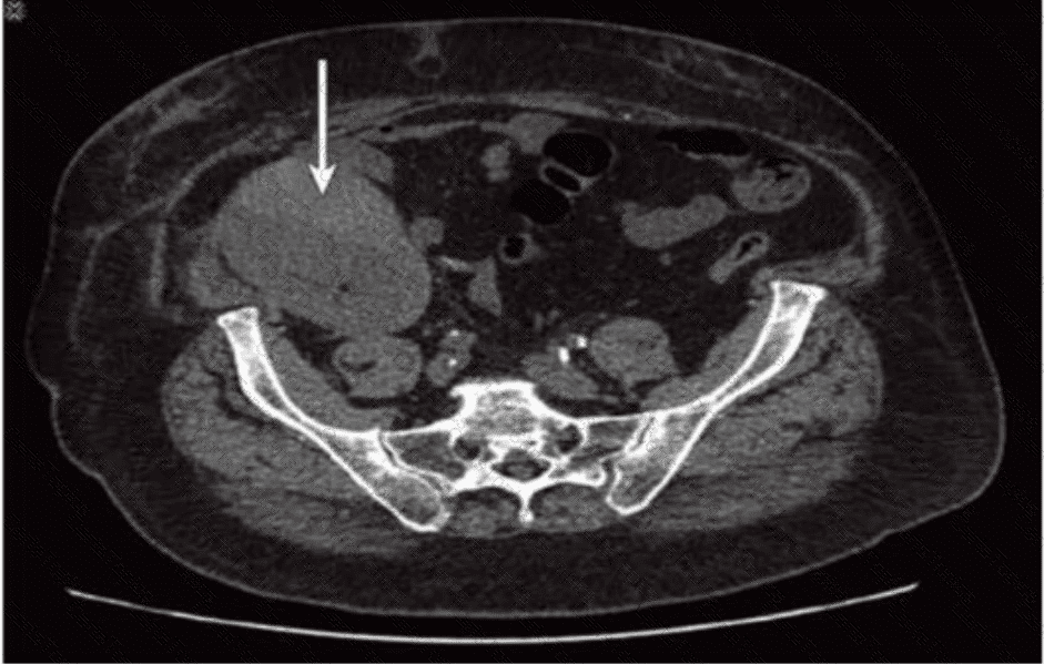
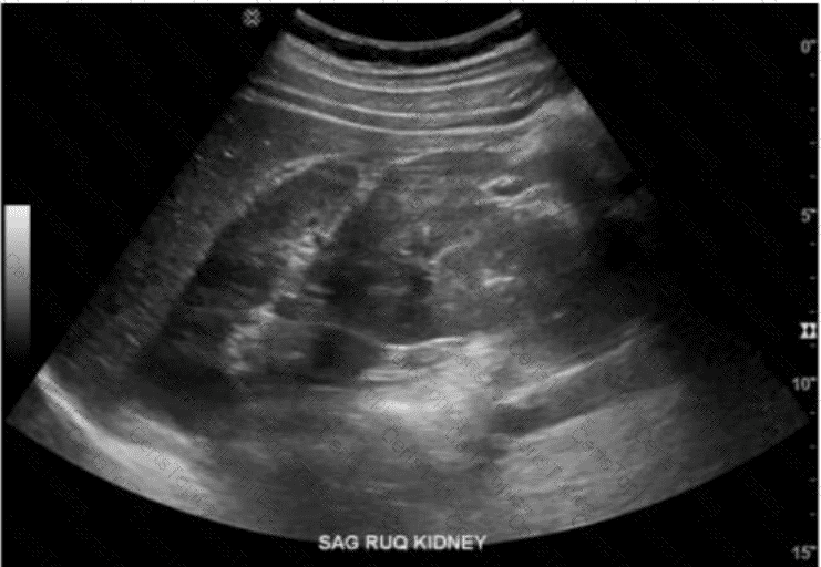
Which condition of the transplant kidney is indicated by the arrows on these images?

Options:

A.

Urinoma

B.

Lymphocele

C.

Renal cell carcinoma

D.

Subcapsular hematoma

Question 22

Which of the following is a possible early complication of a renal transplant?

Options:

A.

Ureterocele

B.

Acute tubular necrosis

C.

Transplant artery stenosis

D.

Transitional cell carcinoma

Question 23

Which vascular condition is most consistent with patent cutaneous para-umbilical channels and portal hypertension?

Options:

A.

Caput medusae

B.

Esophageal varices

C.

Coronary vein varices

D.

Splenic vein varices

Question 24

Which of the following is the most common symptom of cholelithiasis?

Options:

A.

Jaundice

B.

Flank pain

C.

Biliary colic

D.

Hemoptysis

Question 25

Elevation of alpha-fetoprotein levels is a characteristic finding in which tumor?

Options:

A.

Hepatoma

B.

Adenoma

C.

Cholangiocarcinoma

D.

Focal nodular hyperplasia

Question 26

Where is the most common location for a branchial cyst in relation to the thyroid?

Options:

A.

Lateral

B.

Medial

C.

Anterior

D.

Posterior

Question 27

Which gray scale artifact is caused by the oscillation of gas bubbles?

Options:

A.

Reverberation

B.

Mirror image

C.

Ring down

D.

Refraction

Question 28

Which technique may provide better visualization of the common bile duct in a patient with hepatic steatosis?

Options:

A.

Decrease overall gain

B.

Increase dynamic range

C.

Scan patient after a fatty meal

D.

Decrease transducer frequency

Question 29

Which abnormality is the most common adult adrenal tumor?

Options:

A.

Neuroblastoma

B.

Adrenal cortical carcinoma

C.

Pheochromocytoma

D.

Adenoma

Question 30

What is the most common cause of nutcracker syndrome?

Options:

A.

Compression of left renal vein between inferior vena cava and aorta

B.

Compression of left renal vein between superior mesenteric artery and aorta

C.

Compression of right renal vein between superior mesenteric artery and aorta

D.

Compression of right renal vein between inferior vena cava and aorta

Question 31

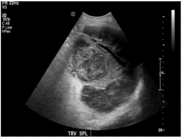
Which clinical finding is most likely associated with the splenic pathology demonstrated in this image?

Options:

A.

Trauma

B.

Sickle cell anemia

C.

Immunocompromised

D.

Portal hypertension

Question 32

Which parameter is most likely increased distal to a renal artery stenosis?

Options:

A.

Resistive index

B.

Pulsatility index

C.

Acceleration time

D.

Spectral broadening

Question 33

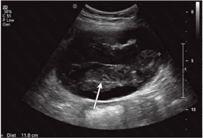
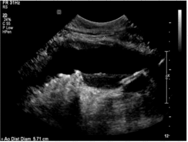
Which sonographic finding is most consistent with this image of the abdominal aorta?

Options:

A.

Stenosis

B.

Dissection

C.

Aneurysm

D.

Occlusion

Question 34

Which sonographic appearance of the bile ducts is demonstrated in this image?

Options:

A.

Normal intrahepatic

B.

Dilated intrahepatic

C.

Dilated common bile

D.

Dilated common hepatic

Question 35

Which renal anomaly is demonstrated on this image?

Options:

A.

Duplicated collecting system

B.

Crossed renal ectopia

C.

Horseshoe kidney

D.

Pelvic kidney

Question 36

Which sonographic appearance of the normal epididymis is the most common?

Options:

A.

Isoechoic to the testis

B.

Anechoic with hyperechoic borders

C.

Homogeneous compared to the testis

D.

Hypoechoic with irregular borders

Question 37

A patient presents with ampulla of Vater obstruction, distention of the gallbladder, and painless jaundice. Which condition is most likely associated with these findings?

Options:

A.

Porcelain gallbladder

B.

Mirizzi syndrome

C.

Courvoisier sign

D.

Choledochal cyst

Question 38

Where in the neck are most thyroid cancer recurrences found?

Options:

A.

Contralateral

B.

Ipsilateral

C.

Bilateral

D.

Subauricular

Question 39

Which imaging technique best demonstrates ureteral patency?

Options:

A.

Spectral Doppler

B.

Gray scale

C.

Color Doppler

D.

Graded compression

Question 40

Which action should a sonographer take if the abdominal aorta measures 5.5 centimeters in the anteroposterior diameter?

Options:

A.

Report the finding to the radiologist immediately

B.

Follow the routine protocol for abdominal ultrasound

C.

Release patient from care

D.

Disclose the diagnosis to the patient

Question 41

Which pancreatic condition is commonly associated with complete or partial atresia of the duodenum?

Options:

A.

Pancreatic cysts

B.

Pancreas divisum

C.

Pancreatic agenesis

D.

Annular pancreas

Question 42

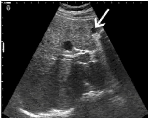
Which retroperitoneal finding is most likely associated with trauma?

Options:

A.

Neuroblastoma

B.

Fibrosis

C.

Urinoma

D.

Adenoma

Question 43

Which sonographic feature is typical of a thyroid adenoma?

Options:

A.

Irregular border

B.

Hypoechoic halo

C.

Hyperechoic nodule

D.

Through transmission

Question 44

Which foreign body is better visualized with sonography than computed tomography (CT)?

Options:

A.

Glass

B.

Wood

C.

Metal

D.

Stone

Question 45

Which condition puts the patient at greatest risk for a hematoma as a result of biopsy?

Options:

A.

Infection

B.

Hypertension

C.

Liver disease

D.

Acute renal failure

Question 46

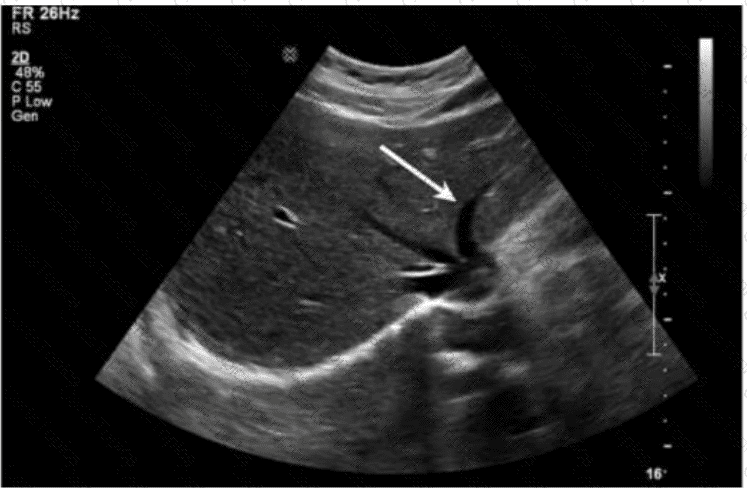
Which structure is indicated by the arrow on this image?

Options:

A.

Left portal vein

B.

Middle hepatic vein

C.

Proper hepatic artery

D.

Inferior vena cava

Question 47

Which condition is demonstrated in this image?

Options:

A.

Intussusception

B.

Pyloric stenosis

C.

Hydronephrosis

D.

Gastritis

Question 48

Which cause of transudative pleural effusion is most common?

Options:

A.

Congestive heart failure

B.

Pulmonary emboli

C.

Lymphoma

D.

Empyema

Question 49

Which condition is most likely associated with this image of the common bile duct?

Options:

A.

Liver mass

B.

Cystic duct stone

C.

Pancreatic head mass

D.

Gallbladder stones

Page: 1 / 12
Total 165 questions